Women Imaging
Women Imaging
We are committed to offering a comprehensive approach to breast health. Our breast-imaging center is at the forefront of this initiative and provides diagnostic radiology services, which includes:
- Screening and diagnostic mammograms
- Breast ultrasound
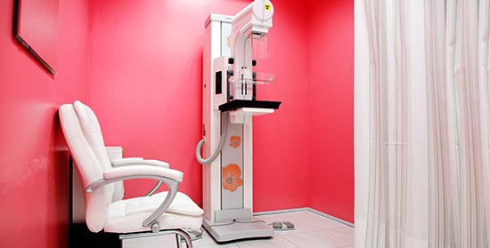
How Often Should You Get A Mammogram? The American Cancer Society recommends a yearly mammogram for all women, age 40 and older, as well as those who may be at higher risk for breast cancer (e.g. family history). This routine screening mammogram takes approximately 15 minutes to performand usually includes capturing two radiographic images of each breast. Mammography Saves Lives. A screening mammogram helps detect breast cancer in the early stages, before it has spread. On the other hand, a diagnostic mammogram provides a more detailed X-ray of the breast. It is used to further investigate suspicious changes found on a screening mammogram. A diagnostic mammogram may be needed if there is:
- Breast pain
- Nipple discharge
- Skin thickening
- A lump
- Changes in breast size or shape
Breast Ultrasound
An ultrasound is often used to supplement diagnostic mammograms in order to evaluate palpable findings, masses, cysts, or ductal abnormalities. Ultrasound is particularly helpful to identify whether a mammographic finding is solid or filled with fluid (cyst). Ultrasound is not appropriate for screening, as it does not detect calcifications, which can be seen in certain types of cancer. During the procedure:
- You will lie down for the exam
- You will keep your arm above your head
- The sonographer will place some gel on your skin and press firmly with the transducer on your breast over the area of interest to obtain images
Images are reviewed in real time by the radiologist on site. If a suspicious finding is identified, you and your referring doctor will be informed at the time of the exam.